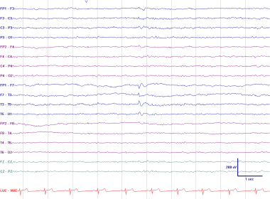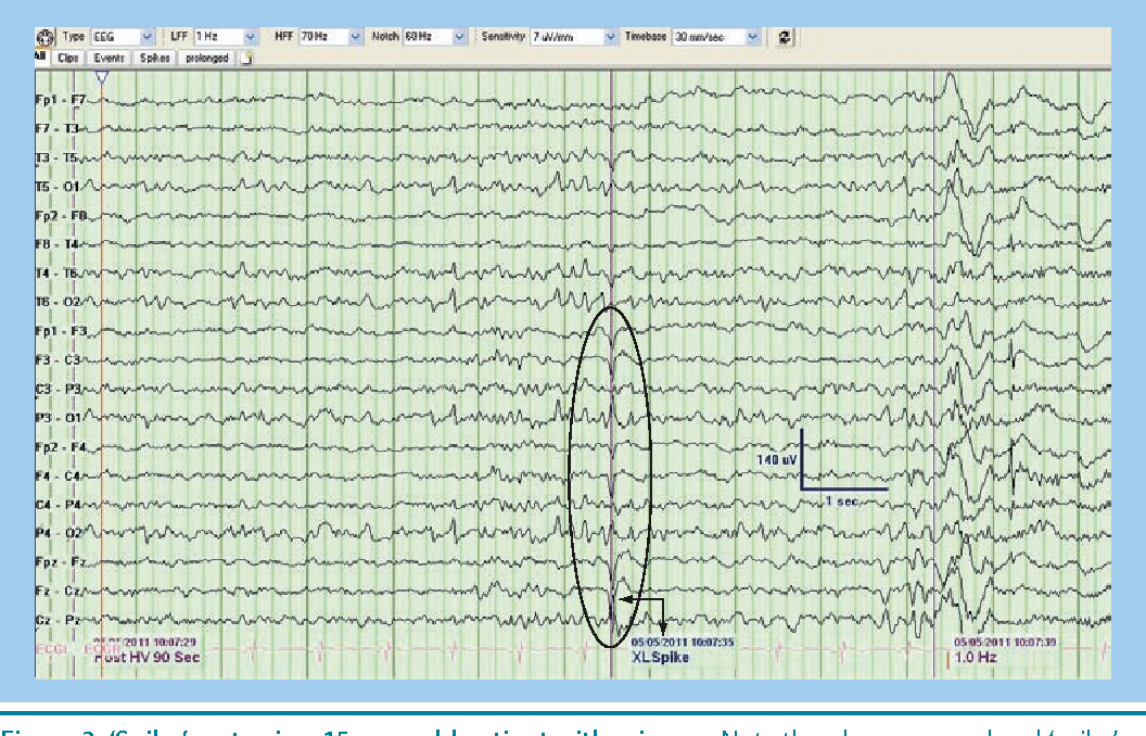


However, in certain comatose states, there can be diffuse alpha activity (alpha comma) and may be considered pathognomonic. For example, alpha waves are seen over the posterior head regions in a normal awake person and considered as the posterior background rhythm. The electroencephalographer is expected to have the significant skills to recognize artifacts, and also an understanding of normal, benign variants. This article reviews the abnormal waveforms in EEG recordings.Įven normal EEG waveforms can be considered potentially abnormal, depending upon various factors. Deep electrical activity of the brain is not well sampled in an EEG using extracranial electrode monitoring.Ībnormal waveforms seen in an EEG recording include epileptiform and non-epileptiform abnormalities. In order to identify abnormal waveforms in EEG, the reader should have a basic understanding of the normal EEG pattern in various physiological states in children and adults. It represents fluctuating dendritic potentials from superficial cortical layers, which are recorded in an organized array pattern and require voltage amplification to be captured. It is a tracing of voltage fluctuations versus time recorded from multiple electrodes placed over the scalp in a specific pattern to sample different cortical regions.

Download the image for free here.Electroencephalography (EEG) was first used in humans by Hans Berger in 1924. Licensed under a Creative Commons Attribution 4.0 International License. At point B, a mix of excitatory and inhibitory postsynaptic potentials result in a different result for the membrane potential. At point A, several different excitatory postsynaptic potentials (EPSPs) add up to a large depolarization. It takes a minimum area of 6 cm² of cortical activation to produce recordable epileptic discharges on standard scalp EEG.įigure 1: Postsynaptic Potential Summation The result of the summation of postsynaptic potentials is the overall change in the membrane potential.The region of the brain with the lowest threshold to electrical stimulation is the hippocampus.It measures a summation of postsynaptic potentials. The EEG does not measure single action potentials.Pyramidal neurons contribute to the plurality of the signal (Figure 1). The electrical fields that generate EEG signals are the result of inhibitory and excitatory postsynaptic potentials (IPSPs and EPSPs) on the apical dendrites of cortical neurons.EEG measures the potential difference between two electrodes on the scalp.


 0 kommentar(er)
